

Granular cell dermatofibroma: a potential diagnostic pitfall
All claims expressed in this article are solely those of the authors and do not necessarily represent those of their affiliated organizations, or those of the publisher, the editors and the reviewers. Any product that may be evaluated in this article or claim that may be made by its manufacturer is not guaranteed or endorsed by the publisher.
Authors
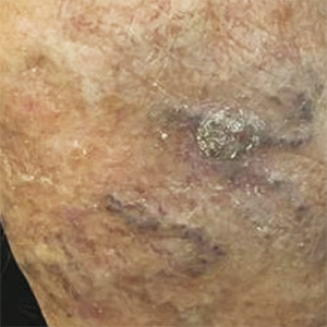
Dermatofibroma, also known as fibrous histiocytoma, is one of the most common cutaneous soft-tissue tumours. Many variants of dermatofibromas have been described and knowledge of these variations is important to avoid a misdiagnosis of a possibly more aggressive tumor. Histological features of different variants can coexist in the same lesion, but typical common fibrous histiocytoma features are generally found, at least focally, in all cases. However, when cellular changes make up the majority of the lesion, the histopathological diagnosis can become more complex and requires immunohistochemical investigations for a correct nosographic classification. We report on the case of a cutaneous fibrous histiocytoma, “granular cell” variant, found on the left leg of a 74-year old woman.
Altmetrics
Downloads
Citations


How to Cite

This work is licensed under a Creative Commons Attribution-NonCommercial 4.0 International License.