

Primary cutaneous adenoid cystic carcinoma of the scalp: dermatosurgical approach with favourable outcome
All claims expressed in this article are solely those of the authors and do not necessarily represent those of their affiliated organizations, or those of the publisher, the editors and the reviewers. Any product that may be evaluated in this article or claim that may be made by its manufacturer is not guaranteed or endorsed by the publisher.
Authors
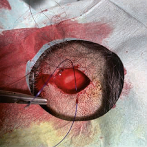
Although described as early as 1975 as a distinct, rare form of cancer with diverse localization, primary cutaneous adenoid cystic carcinoma (PCACC) remains a mystery and challenge for both clinicians and pathologists. The clinical presentation cannot be clearly distinguished from amelanotic melanoma or intradermal nevus, Merkel cell carcinoma, trichofolliculoma, trichoepithelioma or other rare tumors of the adnexa, or dermatofibrosarcoma protuber-ans. The histopathological diagnosis requires not only careful evaluation of standard hematoxylin/eosin preparations, but also immunohistochemical staining with a number of markers such as epithelial membrane antigen (EMA), S-100, SOX-10, Ki-67, CD-117 (c-kit), Vimentin, carcinoembryonic antigen (CEA), Ber-EP4 and many others. The surgical approach should consist of excision with margins between 1 and 2 cm, with the choice of margins depending upon the histopathological findings in the primary excisional specimen. We present a 31-year-old patient with an enlarging, ame-lanotic, plaque-like tumor of the scalp with a duration of no more than 18-24 months. Surgical treatment was performed within two surgical sessions with a total resection field of 1.3 cm. A good cosmetic result was achieved.
Altmetrics
Downloads
Citations


How to Cite

This work is licensed under a Creative Commons Attribution-NonCommercial 4.0 International License.