
Wickham striae on skin appendages: a helpful dermoscopic feature
All claims expressed in this article are solely those of the authors and do not necessarily represent those of their affiliated organizations, or those of the publisher, the editors and the reviewers. Any product that may be evaluated in this article or claim that may be made by its manufacturer is not guaranteed or endorsed by the publisher.
Authors
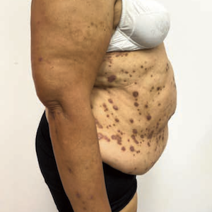
Lichen planus (LP) is a chronic inflammatory disease, clinically characterized by purpuric, itchy papules that typically spread on the trunk and extremities. Other body sites can also be affected, including mucosal membranes, nails, and the scalp. The use of dermoscopy is essential in determining the diagnosis of LP, as it may highlight pathognomonic features such as Wickham striae (WS). WS are thin, pearly white structures arranged in a reticular pattern that is observed over LP lesions and histologically correspond to epidermal hypergranulosis. WS is usually most visible on the oral mucosa but can also cover almost every active LP papule. Here, we report two cases of biopsy-proven LP that show WS on dermoscopy in two specific sites: the scalp and proximal nail fold.
Altmetrics
Downloads
Citations


How to Cite

This work is licensed under a Creative Commons Attribution-NonCommercial 4.0 International License.