Original Articles

Stage-specific tumor microenvironment dynamics and cancer-associated fibroblast profiling in MBL-6 mouse models of breast cancer

Publisher's note
All claims expressed in this article are solely those of the authors and do not necessarily represent those of their affiliated organizations, or those of the publisher, the editors and the reviewers. Any product that may be evaluated in this article or claim that may be made by its manufacturer is not guaranteed or endorsed by the publisher.
Published: 10 October 2025
26
Views
20
Downloads

Authors

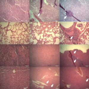
Breast cancer remains the most prevalent malignancy among women, necessitating the development of novel therapeutic strategies. Experimental animal models that closely mimic human breast cancer are crucial for advancing these therapies. This study utilized the criteria of the tumour, node, metastasis (TNM) staging system and variations in metabolic rates to develop models representing stages II and IV of human breast cancer, using the MBL-6 mouse breast cancer cell line. We assessed tumor growth curves in vivo and investigated distant metastasis to organs such as the liver, lungs, lymph nodes, and spleen. Carcinoma-associated fibroblasts (CAFs) were isolated, and their proliferation rates, inflammatory enzyme expression, and matrix metalloproteinase levels were compared between stages II and IV. By analyzing tumor kinetics and metabolic differences, we were able to predict tumor size and progression at each stage. Our results revealed that CAFs isolated from both stages exhibited similar phenotypic characteristics. However, CAFs from stage II tumors showed higher expression of indoleamine 2,3-dioxygenase 1 (IDO1), while those from stage IV tumors had higher levels of inducible nitric oxide synthase (iNOS). These distinct expression patterns suggest unique microenvironmental features at different stages of tumor progression. Further investigation of the cancer microenvironment may provide valuable insights for selecting targeted therapies and improving disease management.

Downloads

Download data is not yet available.

Citations

1. Xu Y, Gong M, Wang Y, et al. Global trends and forecasts of breast cancer incidence and deaths. Sci Data 2023;10:334. DOI: https://doi.org/10.1038/s41597-023-02253-5
2. Jonkers J, Derksen PWB. Modeling metastatic breast cancer in mice. J Mammary Gland Biol Neoplasia 2007;12:191-203. DOI: https://doi.org/10.1007/s10911-007-9050-8
3. Lindsay SJ, Rahbari R, Kaplanis J, et al. Similarities and differences in patterns of germline mutation between mice and humans. Nat Commun 2019;10:4053. DOI: https://doi.org/10.1038/s41467-019-12023-w
4. de Jong M, Maina T. Of mice and humans: are they the same?–Implications in cancer translational research. J Nucl Med 2010;51:501-4. DOI: https://doi.org/10.2967/jnumed.109.065706
5. Terpstra AHM. Differences between humans and mice in efficacy of the body fat lowering effect of conjugated linoleic acid: role of metabolic rate. J Nutr 2001;131:2067-8. DOI: https://doi.org/10.1093/jn/131.7.2067
6. Demetrius L. Of mice and men: when it comes to studying ageing and the means to slow it down, mice are not just small humans. EMBO Rep 2005;6:S39-44. DOI: https://doi.org/10.1038/sj.embor.7400422
7. Hansen K, Khanna C. Spontaneous and genetically engineered animal models; use in preclinical cancer drug development. Eur J Cancer 2004;40:858-80. DOI: https://doi.org/10.1016/j.ejca.2003.11.031
8. Setlow RB. Human cancer: etiologic agents/dose responses/DNA repair/cellular and animal models. Mutat Res 2001;477:1-6. DOI: https://doi.org/10.1016/S0027-5107(01)00090-2
9. Yang Y, Yang HH, Hu Y, et al. Immunocompetent mouse allograft models for development of therapies to target breast cancer metastasis. Oncotarget 2017;8:30621. DOI: https://doi.org/10.18632/oncotarget.15695
10. Ruggeri BA, Camp F, Miknyoczki S. Animal models of disease: pre-clinical animal models of cancer and their applications and utility in drug discovery. Biochem Pharmacol 2014;87:150-61. DOI: https://doi.org/10.1016/j.bcp.2013.06.020
11. Szadvári I, Krizanova O, Babula P. Athymic nude mice as an experimental model for cancer treatment. Physiol Res 2016;65. DOI: https://doi.org/10.33549/physiolres.933526
12. Olson B, Li Y, Lin Y, et al. Mouse models for cancer immunotherapy research. Cancer Discov 2018;8:1358-65. DOI: https://doi.org/10.1158/2159-8290.CD-18-0044
13. Karamitopoulou E. Tumour microenvironment of pancreatic cancer: immune landscape is dictated by molecular and histopathological features. Br J Cancer 2019;121:5-14. DOI: https://doi.org/10.1038/s41416-019-0479-5
14. Langroudi L, Hasan ZM, Ardeshirylajimi A, Soleimani M. Isolation and characterization of a new cell line from spontaneous mouse mammary tumour, MBL-6, for in vivo cancer studies. Vet Sci Dev 2017;7. DOI: https://doi.org/10.4081/vsd.2015.6042
15. Edelstein-Keshet L. Mathematical models in biology. SIAM; 2005. DOI: https://doi.org/10.1137/1.9780898719147
16. Amin MB, Greene FL, Edge SB, et al. The Eighth Edition AJCC Cancer Staging Manual: Continuing to build a bridge from a population-based to a more "personalized" approach to cancer staging. CA Cancer J Clin 2017;67:93-9. DOI: https://doi.org/10.3322/caac.21388
17. Faustino-Rocha A, Oliveira PA, Pinho-Oliveira J, et al. Estimation of rat mammary tumor volume using caliper and ultrasonography measurements. Lab Anim (NY) 2013;42:217-24. DOI: https://doi.org/10.1038/laban.254
18. Yang CH, Wang SJ, Lin ATL, et al. Evaluation of prostate volume by transabdominal ultrasonography with modified ellipsoid formula at different stages of benign prostatic hyperplasia. Ultrasound Med Biol 2011;37:331-7. DOI: https://doi.org/10.1016/j.ultrasmedbio.2010.10.026
19. Alimohamadi Y, Sepandi M. Sample Size in Animal Studies (The number of laboratory animals in a Research study). Iran J Med Microbiol 2022;16. DOI: https://doi.org/10.30699/ijmm.16.2.173
20. Lavie D, Ben-Shmuel A, Erez N, Scherz-Shouval R. Cancer-associated fibroblasts in the single-cell era. Nat Cancer 2022;3:793-807. DOI: https://doi.org/10.1038/s43018-022-00411-z
21. Lemos H, Huang L, Prendergast GC, Mellor AL. Immune control by amino acid catabolism during tumorigenesis and therapy. Nat Rev Cancer 2019;19:162-75. DOI: https://doi.org/10.1038/s41568-019-0106-z
22. Bandeira LG, Bortolot BS, Cecatto MJ, et al. Exogenous tryptophan promotes cutaneous wound healing of chronically stressed mice through inhibition of TNF-α and IDO activation. PLoS One 2015;10:e0128439. DOI: https://doi.org/10.1371/journal.pone.0128439
23. Hornyák L, Dobos N, Koncz G, et al. The role of indoleamine-2, 3-dioxygenase in cancer development, diagnostics, and therapy. Front Immunol 2018;9:340144. DOI: https://doi.org/10.3389/fimmu.2018.00151
24. Deng J, Li D, Huang X, et al. Interferon-γ enhances the immunosuppressive ability of canine bone marrow-derived mesenchymal stem cells by activating the TLR3-dependent IDO/kynurenine pathway. Mol Biol Rep 2022;49:8337-47. DOI: https://doi.org/10.1007/s11033-022-07648-y
25. Braun D, Longman RS, Albert ML. A two-step induction of indoleamine 2, 3 dioxygenase (IDO) activity during dendritic-cell maturation. Blood 2005;106:2375-81. DOI: https://doi.org/10.1182/blood-2005-03-0979
26. Cheng JT, Deng YN, Yi HM, et al. Hepatic carcinoma-associated fibroblasts induce IDO-producing regulatory dendritic cells through IL-6-mediated STAT3 activation. Oncogenesis 2016;5:e198. DOI: https://doi.org/10.1038/oncsis.2016.7
27. Biswas P, Stuehr DJ. Indoleamine dioxygenase and tryptophan dioxygenase activities are regulated through control of cell heme allocation by nitric oxide. J Biol Chem 2023;299. DOI: https://doi.org/10.1016/j.jbc.2023.104753
28. Ye QX, Xu LH, Shi PJ, et al. Indoleamine 2, 3-dioxygenase and inducible nitric oxide synthase mediate immune tolerance induced by CTLA4Ig and anti-CD154 hematopoietic stem cell transplantation in a sensitized mouse model. Exp Ther Med 2017;14:1884-91. DOI: https://doi.org/10.3892/etm.2017.4722
29. Vafaei S, Taheri H, Hajimomeni Y, et al. The role of NLRP3 inflammasome in colorectal cancer: Potential therapeutic target. Clin Transl Oncol 2022;24:1881-9. DOI: https://doi.org/10.1007/s12094-022-02861-4

Supporting Agencies

This study was funded by the Research Deputy of Tarbiat Modares University.

How to Cite


Stage-specific tumor microenvironment dynamics and cancer-associated fibroblast profiling in MBL-6 mouse models of breast cancer. (2025). Veterinary Science Development, 9(1). https://doi.org/10.4081/vsd.2025.10069