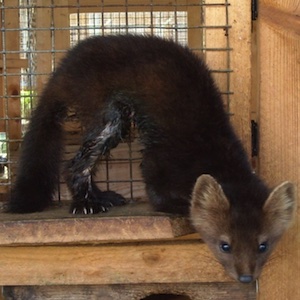
Pathological findings in ‘wet belly’ in young black sables (Martes zebellina)
All claims expressed in this article are solely those of the authors and do not necessarily represent those of their affiliated organizations, or those of the publisher, the editors and the reviewers. Any product that may be evaluated in this article or claim that may be made by its manufacturer is not guaranteed or endorsed by the publisher.
Accepted: 22 May 2025
Authors
‘Wet belly’ in industrial sables is an economically significant problem of modern industrial fur farming in Russia due to defects in the skin and fur of animals, which are associated with the damaging effect of constantly excreted urine. The incidence in young sables in different years can range from 0.1 to 6% in this age group. In this regard, in case of ‘wet belly’ disease in young sables, the regulations of veterinary intervention provide only local aerosol treatment with antimicrobials.
There are practically no studies devoted to the search for the causes of wet belly disease in young sables, including from the point of view of analyzing the results of autopsy. The causes and pathophysiological mechanisms of ‘wet belly’ development remain unknown, in particular, the mechanisms of urinary disorders and urinary incontinence. The present article is the first attempt to analyze the autopsy data of 76 young sables, which showed that quite a large number of animals had concomitant pathology of the digestive and respiratory system, as well as clinical and histological signs of cystitis and probable urinary tract infection (UTI).
Downloads
Citations


Supporting Agencies
This work was supported by the Sechenov Institute of Evolutionary Physiology and Biochemistry of the Russian Academy of Sciences (State assignment No. 075-00264-25-00).How to Cite

This work is licensed under a Creative Commons Attribution-NonCommercial 4.0 International License.